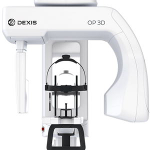
DEXIS i-CAT™ FLX V-SERIES
Cone Beam 3D Imaging.The i-CAT FLX V-Series offers three maximum fields of view (FOV): V8, V10, and V17. Within these sizes, you have the option to scale or collimate the scan height to capture only the area of interest per the patient’s immediate need. The system also provides enhanced low-dose imaging options, dedicated traditional 2D panoramic capabilities, and the feature-rich… Read more
Promotions
Description
Cone Beam 3D Imaging.The i-CAT FLX V-Series offers three maximum fields of view (FOV): V8, V10, and V17. Within these sizes, you have the option to scale or collimate the scan height to capture only the area of interest per the patient’s immediate need. The system also provides enhanced low-dose imaging options, dedicated traditional 2D panoramic capabilities, and the feature-rich treatment planning software.
It’s hard to choose when all three models have class-leading performance.
i-CAT FLX V8 – With 8 cm x 5 cm to 8 cm x 8 cm FOVs, this system accommodates those dentists
who want to capture select treatment areas for survey, implant, oral surgery, and endodontics.
i-CAT FLX V10 – The V10 offers a maximum and scalable FOV of up to 16 cm D x 10 cm H, helpful
when you expand into larger cases and airway or TMJ analysis.
i-CAT FLX V17 – With a maximum FOV of up to 23 cm D x 17 cm H, the V17 is used most
often by orthodontics, oral maxillofacial surgeons, and oral radiologists.
Low-Dose Options
i-CAT QuickScan and QuickScan+ protocols allow you to take complete 3D images at a radiation dose comparable to a 2D panoramic image*. The flexibility in scalable scan sizes further allows you to limit the dose to the patient.
*Utilizing the i-CAT FLX QuickScan+ exposure tool protocol. Use of lower dosage imaging may only be suitable for certain diagnostic tasks. Image quality is proportional to dose. i-CAT FLX offers a variety of exposure protocols allowing clinicians to adjust dosage to specific diagnostic needs.
Open Platform
i-CAT FLX allows for an open platform when using your DICOM files. These files are not ‘locked’, giving you the freedom to use i-CAT-captured files in a wide array of programs including CAD/CAM applications and other dental equipment.
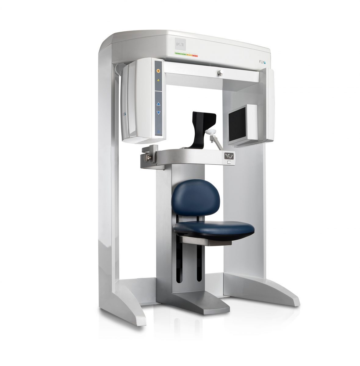
Seated Position
i-CAT FLX is one of the few machines that provides a seated position for scanning. Ergonomic Stability System (ESS) allows for easy, seated patient positioning, designed to minimize patient movement, and avoid unnecessary retakes and additional radiation.
Traditional 2D Panoramics
Your “2-in-1” i-CAT FLX V-Series takes a traditional 2D pan using the same high-quality sensor that is used to acquire 3D scans. Using your system to capture BOTH 2D and 3D imaging assists you with your ALARA goals and helps you transition to 3D imaging at your own pace.
Full 24.2 cm x 19.3 cm Sensor
All i-CAT FLX V-Series models come equipped with a full 24.2 cm x 19.3 cm sensor capable of yielding a full ceph height 3D scan (13 cm x 16 cm) in just 4.8 seconds. This allows owners the flexible freedom to upgrade their field-of-view without the need to replace the unit itself. The solution is simply an in-office software upgrade that opens the field-of-view to the next V-Series configuration.
No Offset Scanning
With our PureScan™ technology and full-size sensor, i-CAT FLX V-Series scanners capture a 3D scan without compromising image quality. This full-beam scanning maximizes the use of the large sensor to provide more anatomical information at a higher quality compared to other smaller sensors.
Unlike the i-CAT FLX V-Series, some 3D CBCT imaging machines use a smaller sensor, which only captures half of the volume at a time requiring images to be stitched together. Offset or half-beam scanning allows a smaller sensor to shift and scan a slightly larger volume than its size, although the cost associated is the sacrifice of image quality and anatomical accuracy.
Clinical Indications
Your dental practice is unique—that’s why you need a flexible 3D solution that works with your practice and provides the control you need to deliver optimum care to your patients.
The i-CAT FLX V-series offers effective treatment planning and predictability to help general dentists and specialists alike by providing fast sizable scans, low-dose options, and dedicated specialty tools within Tx STUDIO, powered by Anatomage.
Implantology
Using the i-CAT FLX V8, V10 or V17, clinicians can place and restore with accuracy and confidence.
Treat patients with greater surgical predictability and confident outcomes using i-CAT’s 3D treatment planning tools. Use i-CAT’s high resolution, volumetric images and complete 3D views for a more thorough analysis of bone structure and tooth orientation. Collect precise data and map an entire course of treatment for surgical placement of the implant and abutment, all the way to final restoration.
Oral and Maxillofacial Surgery
Tx STUDIO treatment planning software, powered by Anatomage, can assist in identifying deformities, such as cysts, tumors, lesions, and changes of the jaw, to avoid potential surgical complications. Determine precise position of impacted teeth within the alveolar bone, as well as their proximity to adjacent teeth and vital structures, such as the nerve canal, sinus walls, and cortical borders.
Using the i-CAT FLX V8, V10 or V17, clinicians can place and restore with accuracy and confidence.
Treat patients with greater surgical predictability and confident outcomes using i-CAT’s 3D treatment planning tools.
Use i-CAT’s high resolution, volumetric images and complete 3D views for a more thorough analysis of bone structure and tooth orientation.
Collect precise data and map an entire course of treatment for surgical placement of the implant and abutment, all the way to final restoration.
Orthodontics
With the I-CAT FLX V17, clinicians can understand exact tooth position and the relationship of anatomy so they can map the most effective, least invasive treatment plan for the best alignment. Correct root angulations and find supernumerary teeth and their exact locations to enhance communication with oral surgeons. Additional treatment modules in the 3D treatment planning software, including 3D cephalometric analysis, virtual studies and impressionless models, make planning even more powerful.
Enhanced communication with oral surgeons
Clinicians can utilize the image data provided by i-CAT FLX V17 and the 3D treatment planning software to correct root angulations and find exact locations of supernumerary teeth. Exploratory surgery may be prevented based upon the clinician’s better understanding of the patient’s conditions.
Additional Treatment Modules
Planning is even more powerful with 3D cephalometric analysis, virtual studies, and impression-less models. Incorporate CBCT scans in digital orthodontic treatment plans and personalized appliances.
Endodontics
Using the i-CAT FLX V8, V10, or V17, clinicians can survey roots in three dimensions.
When more concentrated studies are necessary, high resolution scans – up to .125 voxels – lend more detail for the identification of lesions. Scans can also be collimated to cover the area of interest.
Within Tx STUDIO software, scans can be explored axially and buccolingually for a complete survey of fractures, accessory canals and endo-perio involvement.
Airway Analysis
Using the i-CAT FLX V10 or V17, clinicians can evaluate airway obstructions to help identify patients with an increased risk of a possible sleep disorder.
For patients with suspect airway or sinus tissues, you can use Tx STUDIO software to review the 3D data and to reveal restricted airways and determine appropriate treatments with precise anatomical views and measurements. Assess airway volume-at-a-glance using color-coded constriction values. Quickly trace airway on-screen to perform automatic calculations and measurements of paranasal sinuses to evaluate treatment options. i-CAT is FDA cleared to provide sufficient and appropriate data from 3D volumes of the maxillofacial anatomical structures, including the airway.
Periodontics
Using the i-CAT FLX V10 or V17, clinicians can analyze bone structure.
From implant placement to surgical options for the management of bone loss, the i-CAT FLX provides periodontists with a wide range of services expected from the specialty. Capture 3D volumetric images for a more thorough analysis of bone structure as well as sinus and nerve location.
i-CAT imaging system provides options and a full complement of tools for implant placement. Use scan data to help plan the course of treatment of bony defects prior to the actual osseous surgery appointment. With Tx STUDIO software’s highly visual 3D presentation, share your diagnosis and plan with the patient for greater understanding leading to better post-treatment compliance.
TMJ
Using the i-CAT FLX V10 or V17, clinicians can detect TMD and access fractures.Detect TMJ anomalies for the ability to design effective patient treatment. Using the TMJ visualization tools, zero in on the temporomandibular joints to identify wear, defects, and fractures. These tools also act as virtual study models to streamline and expedite treatment and allow you to design splints with the optional Medical Design Studio module.
Specifications
Sensor Type: Amorphous Silicon Flat Panel Sensor with Csl Scintillator
Grayscale Resolution: 16-bit
Voxel Size: .4 mm, .3 mm, .25 mm, .2 mm, .125 mm
Collimation: Electronically controlled fully adjustable collimation
Scan Time: 4.8, 8.9, 14.7, 17.8 or 26.9 seconds
Exposure Type: Pulsed
Field-of-View:
- V8: 8cm x 5cm, 8cm x 8cm
- V10: 8cm x 5cm, 8cm x 8cm, 16cm x 4cm, 16cm x 6cm, 16cm x 8cm, 16cm x 10cm
- V17: 8cm x 5cm, 8cm x 8cm, 16cm x 4cm, 16cm x 6cm, 16cm x 8cm, 16cm x 10cm, 16cm x 11cm, 16cm x 13cm, 23cm x 17cm
Reconstruction Shape: Cylinder
Typical Reconstruction Time: Less than 30 seconds
Viewing and Treatment Software: Included
DICOM Compatible: Yes
Unit Size: 48″ (w) x 69.5″ (h) x 36.37″ (d)
Patient Position: Seated
Wheelchair Accessible: Yes
Technical Drawings