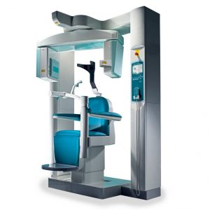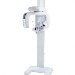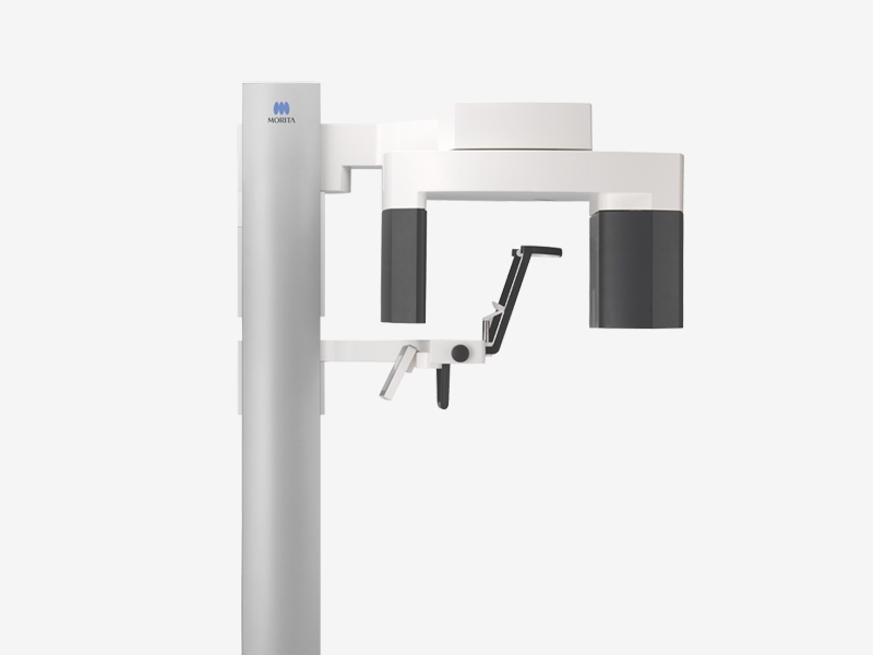

J. Morita Veraview X800 3D Panoramic
Revolutionary Image Quality Veraview X800 is a newly redesigned multifunctional X-ray unit loaded with practical, unique, and refined features. Image clarity has been enhanced with a voxel size of just 8 micrometer and a resolution of 2.5 LP/mm, the highest level of any Morita multifunctional unit to date. Veraview X800 offers progressive technology that automatically sets the optimal x-ray beam angle,… Read more
Promotions
Literature
J. Morita Veraview X800 3D Pan Brochure
Description
Revolutionary Image Quality
Veraview X800 is a newly redesigned multifunctional X-ray unit loaded with practical, unique, and refined features. Image clarity has been enhanced with a voxel size of just 8 micrometer and a resolution of 2.5 LP/mm, the highest level of any Morita multifunctional unit to date.
Veraview X800 offers progressive technology that automatically sets the optimal x-ray beam angle, horizontal for 3D or raised 5° for 2D panoramic images. This slight upward projection for panoramic imaging enhances image quality by eliminating potential super imposition of anatomical structures including the shadow from the base of the skull and hard palate over the maxillary teeth. For 3D imaging, a horizontal beam minimizes distortion and reduces metal artifacts. The X800 unit, therefore, self-adjusts to optimize image quality during both acquisition techniques.
Extra-sharp images: The smallest details at a glance –
Sharp imaging plays a key role in patient health. The sharper the image, the more accurate the diagnosis and treatment.
Top performance: Peak values of 80 μm and 2.5 LP/mm
Image clarity is where the Veraview X800 is especially impressive. It produces images of 80 μm for Ø 40 mm × H 40 mm fields of view (FOV) and a brilliant resolution of 2.5 LP/mm.
MTF (Modulation Transfer Function) is one way to objectively evaluate the line pair resolution and objectively express how many line-pairs and what level of contrast can be discriminated. Generally, if MTF is 10%, naked eye discrimination is possible. Spatial resolution does not depend only on voxel size.
Minimal artifacts: Best image quality for the most reliable diagnoses –
For a clear diagnosis, you need a clear X-ray image. Artifacts in an image can mimic pathological alterations that can even lead to false findings. That’s why it’s so important to keep these “false positives” from showing up in the first place, and to smooth the way for an unambiguous, reliable diagnosis.
A clever concept: Horizontal X-ray beam
Veraview X800 is designed to keep artifacts and distortions to a minimum. That’s possible because of the horizontal orientation of the X-ray beam, which produces top-quality CBCT exposures. The horizontal X-ray beam prevents artifacts typical of conventional methods (with a raised beam) from occurring. For panoramic exposures, the horizontal X-ray beam is raised by 5º, which suppresses the interfering shadow of the hard palate.
Sharper image or lower dose: 360º or 180º exposure
If you want to get the greatest possible detail, you can use the 360º mode. Depending on the indication, the 180º mode is also available, which offers a shorter exposure time and a lower radiation dose.
Eleven fields of view: Always a perfect setting –
With eleven fields of view (FOVs), the X-ray system provides the diagnostic confidence needed for planning a successful treatment. Following the ALARA principle (as low as reasonably achievable), you can choose the optimum FOV for your diagnostic issue. So you always have the best possible image of the region of interest – even as you minimize the radiation dose.
New: Sharper reconstruction
Now, for the first time, there’s a 2D/3D imaging system with a function for zoom reconstruction. From an original image with a 125 μm voxel size, you can reconstruct a high-resolution image segment at 80 μm – without retaking another image. (This function, however, is not available for the Ø 150 field of view).
Sharp panoramic images: Brilliant all around –
For diagnostics or dental implants – any successful extensive treatment plan relies on a full overview of the teeth and mandibular bone. The Veraview X800, with its numerous innovative functions, offers brilliant opportunities for excellent contrast and consistent high resolution.
Focus on high resolution: The AFP function
The AFP (Adaptive Focal Point) function analyzes multiple layers of exposures. It checks each region, chooses the optimum panoramic layer, and recomposes them into a new image. The result: Everything in the image from the root apex to the incisor region is in perfect focus.
Real-time dose adjustment: The DDAE function
With the DDAE (Digital Direct Auto Exposure) function, the flat panel detector detects X-ray transparency in real time during an exposure, and then adjusts the amount of X-rays emitted to create images with a significantly improved dynamic range.
The royal road: Combining multiple functions
A number of functions, including AFP, AGS, and AIE-HD, can be combined. The result is images that are consistently in precise focus and clearly show the regions of interest.
Face-to-face design: Communicating on the same level –
One element of treatment that’s often underestimated is communication with the patient – yet it’s the foundation for a trusting doctor-patient relationship and can also make treatment procedures significantly easier. It’s a point we factored in from the outset in designing the Veraview X800.
Easy: Positioning for direct patient contact
Face-to-face positioning makes it easier both to communicate with the patient and to position the laser beam.
Intuitive: A pictogram user interface
The touch panel works intuitively and effortlessly using pictograms.
Reduced dose: Less radiation – more protection –
X-ray exposure has decreased dramatically since digital imaging systems were introduced – but patient health remains a core concern. That’s why we do all we can to keep reducing dose in order to provide the greatest possible protection.
Especially for 2D images, the Veraview X800 scores with many innovative features.
Smart and smaller: Collimated images
When you use the partial panoramic mode for 2D images, the focus is pared down to the essentials. The result: the region of interest is clearly visible, while the surrounding areas get a significantly lower dose.
Gentle to the little ones: Special children’s setting
The children’s setting for 2D images reduces irradiation time within the desired region. But grown-ups benefit too. Depending on how big the patient is, you can set the unit for a large, average, or small patient and thereby optimize the relationship between benefits and patient dose.
Also considered: A choice of three image layer orbits
No patient is exactly like any other – people differ not just in size but in many other ways, including the shape of the dental arch. That’s why the exposure layer for 2D images can be optimally adjusted to the patient’s dental arch. Three types of image layer orbits are available: narrow, standard, and wide.
Well selected: Partial cephalometric images –
For cephalometric exposures, three regions can be selected individually in order to reduce the X-ray exposure for the patient. All the same, increasing tube voltage to 100 kV still allows high quality exposures with a resolution of 96 μm.
Network integration: Well-networked everywhere –
PC or tablet: Images available everywhere
CBCT and 2D images can be displayed on any PC or tablet computer using a conventional web browser without installing any special software, which is convenient and helpful for patient consultation
Specifications
- Order Selection F40 / R100 / F150
- Rating AC 120V 60Hz
- AC 220 / 230 / 240 AC voltage; 50 / 60Hz
- Power Consumption 2.0 kVA
- Weight Approx. 185 kg
- X-ray Tube Voltage 60–100 kV (depending on exposure mode)
- X-ray Tube Current 2–10 mA (depending on exposure mode)
- Nominal Focal Spot 0.5