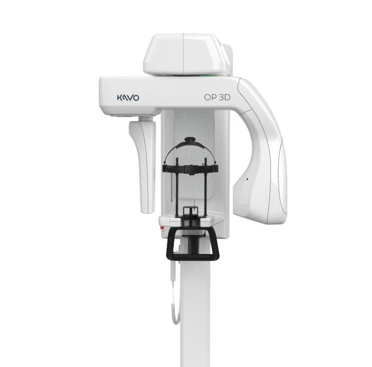
KaVo OP 3D™ upgradable pan
Panoramic imaging with upgrade options to 3D and cephalometric imaging KaVo OP 3D panoramic imaging features a wide variety of views including standard panoramic, pediatric panoramic, TMJ and bitewing. Selecting the image needed is a simple one-click operation. Full upgradability: The OP 3D panoramic unit is completely upgradeable. Choose the addition of cephalometric imaging, or completely upgrade and choose to add… Read more
Promotions
Literature
Description
Panoramic imaging with upgrade options to 3D and cephalometric imaging
KaVo OP 3D panoramic imaging features a wide variety of views including standard panoramic, pediatric panoramic, TMJ and bitewing. Selecting the image needed is a simple one-click operation.
Full upgradability:
The OP 3D panoramic unit is completely upgradeable. Choose the addition of cephalometric imaging, or completely upgrade and choose to add 3D imaging to your practice for even more diagnostic options.
The ORTHOPANTOMOGRAPH™, introduced over 50 years ago, was a revolutionary groundbreaker and pacesetter for dental panoramic X-ray imaging.
Today, with more than 60,000 units sold, the ORTHOPANTOMOGRAPH™ systems are regarded as the leading name and benchmark in the X-ray world.
Excellent Image Quality
With the ORTHOfocusTM feature, the optimum panoramic layer is automatically obtained. This results in easing the patient positioning process and delivering consistent and repeatable image quality even with differences in patient anatomy.
The 9 second standard and pediatric panoramic scan times provide clear image definition, with fewer movement artifacts as well as a lower dose to the patient.
Specifications
3D / CBCT
Image detector: CMOS
Image voxel size: 80–400 μm
Tube voltage: 95 kV
Tube current: 2–12.5 mA
Scan time: 10–20 s
Image volume sizes (H x Ø): 5x 5, 6x 9, 9x 11, 9x 14 cm (optional)
Volume height and location are adjustable through SMARTVIEW™ 2.0 interface.
Wheelchair accessible: Yes
2DPanoramic
Image receptor: CMOS
Pixel size (sensor & image): 99 µm
Tube voltage: 60–90 kV
Tube current: 2–16 mA
Scan time: 9 s
Image field height: 147 mm
Imaging programs: Standard, Segmented, Pediatric, Lat TMJ, Bitewing
2D Cephalometric
Image receptor: CMOS
Pixel size (sensor & image): 99 µm
Tube voltage: 60–95 kV
Tube current: 2–14 mA
Scan time: 10.5 and 8.1 s
Image field height: 180–223 mm
Image field width: 160–260 mm
Imaging programs: Lateral and Pediatric Lateral with an adjustable field width, Posterior- Anterior (PA), Carpus*
Others
Tube focal spot: 0.5 IEC 336 (IEC 60336/2005)
DICOM** support: Available as a software option