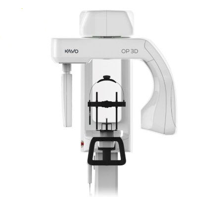
KaVo OP 3D
Your Gateway to 3D The ORTHOPANTOMOGRAPH™ OP 3D unit is designed for advanced dental imaging needs. It is a complete X-ray platform that provides easy-to-use features throughout the entire dental imaging workflow. The versatile panoramic and 3D programs offer imaging excellence for a variety of users ranging from general dental practitioners to maxillofacial surgeons and airway specialists. Designed for efficiency Variety of resolutions… Read more
Promotions
Literature
Description
Your Gateway to 3D
The ORTHOPANTOMOGRAPH™ OP 3D unit is designed for advanced dental imaging needs. It is a complete X-ray platform that provides easy-to-use features throughout the entire dental imaging workflow. The versatile panoramic and 3D programs offer imaging excellence for a variety of users ranging from general dental practitioners to maxillofacial surgeons and airway specialists.
- Designed for efficiency
- Variety of resolutions
- Four versatile fields-of-view (FOV)
- Sustainable green solution
- ORTHOselect™ for optimized workflow
- Customized FOVs with SMARTVIEW™ 2.0
- ORTHOfocus™ for sharp images
- QUICKcompose™ feature for fast image review
- Clearer images with MAR technology
Designed for Efficiency
Every feature of the OP 3D is designed to increase practice efficiency. Preparing the unit for a scan is fast with an intuitive patient positioning system and graphical user-interface. Imaging protocols are optimized for practice workflows.
Variety of Resolutions
- Low Dose TechnologyTM scan (LDT) can be utilized in dose-sensitive cases and in control and follow-up scans where patient dose is to be minimized or lower resolution is acceptable.
- Standard resolution scan with optimized patient dose can be used for general diagnostics.
- High resolution scan offers extremely sharp images for more detailed diagnosis.
- Endo resolution scan with 80 μm voxel size specially designed for endodontic applications. Endo resolution is available for the 5 x 5 FOV.
Clinical Applications
3D images provide valuable information vital to diagnosis and optimal treatment planning. Evaluation of different morphologies is easy as the region of interest can be viewed from all directions.
- Implantology
- Endodontics
- Impactions
- TMJ
- Airway
- Trauma
- Periodontics
Sustainable Green Solution
The OP 3D replaces lead typically used for tubehead radiation shielding designs with a more ecological and environmentally friendly alternative that provides equivalent radiation attenuation. Plus, the power save feature of this system reduces overall energy consumption of the practice.
ORTHOselect™ for Optimized Workflow
The desired imaging area can be selected intuitively with the ORTHOselect user interface. Selections can be made as individual teeth, an entire upper or lower jaw, or TMJ. The optimum field-of-view (FOV) is set automatically based on the selection.
Customized FOVs with SMARTVIEW™ 2.0
With OP 3D, the number of FOV sizes is practically unlimited. SMARTVIEW 2.0 user interface enables choosing the most optimum FOV size for the clinical need as the FOV height and width can be freely adjusted from the taken scout image.
ORTHOfocus™ Sharp Images Automatically
With the ORTHOfocus feature, the optimum panoramic image layer is automatically obtained enabling forgiving patient positioning. The result is consistent image quality every time.
QUICKcompose™ for Fast Image Review
QUICKcompose, available for both panoramic and 3D modalities, offers a quick preview of the captured image allowing for timely evaluation.
Specifications
| Focal Spot | 0.5 mm, IEC 336 (IEC 60336/2005) |
| Tube Voltage | 60-95 kV |
| Tube Current | 3.2–16 mA |
2D Panoramic | |
| Image Detector | CMOS |
| Sensor Pixel Size | 99 µm |
| Image Pixel Size | 99 µm |
| Scan/Exposure Time | 9 s |
| Image Field Height | 147 mm |
| Imaging Programs | Standard, Segmented, Pediatric, Lat TMJ, Bitewing |
| Wheelchair Accessibility | Yes |
3D | |
| Image Detector | CMOS |
| Image Voxel Size | 80 µm-400 µm |
| Scan Time | 10–20 s |
| Image Volume Sizes (H xW) | 5 x 5, 6 x 9, 9 x 11, 9 x 14 cm |
| DICOM Support | Yes |
Minimum System Requirements for 3D Acquisition Workstation
| CPU (processor) | Intel® Core™ i5, i7 or Xeon®, 4-cores or more |
| GPU (graphics processing unit) | NVIDIA® Quadro® M2000 4GB or GeForce® GTX 1050 Ti 4GB |
| RAM (memory) | 8 GB or more |
| Storage (hard disk) | 1 TB or more RAID 1 or RAID 5 recommended for data redundancy, plus backup |
| Network | Gigabit Ethernet 1000 Mb/s |
| Operating System | Windows® 10 Pro or Enterprise, 64‑bit Windows 8.1 Pro or Enterprise, 64‑bit Windows 7 Professional, Ultimate or Enterprise, 64‑bit, with SP1 |
| Display | 1920 x 1080 resolution (Full HD) or higher, at least 300 cd/m2 brightness for typical room lighting, native contrast ratio 100:1 or better, 8-bit panel strongly recommended |
| Other | OpenCL 1.1 support DVD‑ROM drive Anti‑virus software |
| Notes | Please refer to software and device installation manuals for detailed requirements |
| Core™, GeForce®, Intel®, Microsoft®, NVIDIA®, Quadro®, Windows® and Xeon® are trademarks of their respective owners. | |
