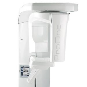
Planmeca ProMax 2D S2
The Extraoral Bitewing Story: Planmeca introduced the most efficient and diagnostic EXTRAORAL bitewings to 2D routine imaging. User interface provides guidance The full-color graphical user interface provides clear texts and symbols to guide you through your procedure. Settings are logically grouped and easy to understand, speeding up imaging and allowing you to focus on your patients. Easy patient positioning Open patient positioning… Read more
Promotions
Literature
Planmeca Accurate Diagnostics, Safer Imaging
Description
The Extraoral Bitewing Story:
Planmeca introduced the most efficient and diagnostic EXTRAORAL bitewings to 2D routine imaging.
User interface provides guidance
The full-color graphical user interface provides clear texts and symbols to guide you through your procedure. Settings are logically grouped and easy to understand, speeding up imaging and allowing you to focus on your patients.
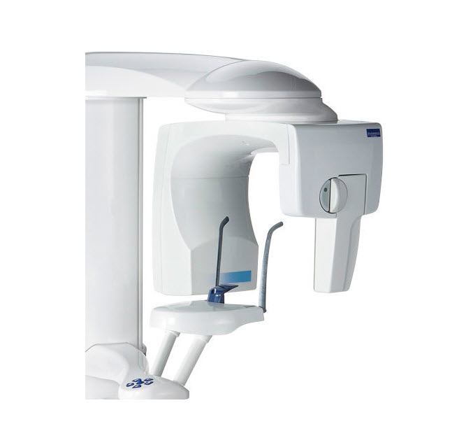
Easy patient positioning
Open patient positioning and side entry minimize errors caused by incorrect patient positioning, allowing you to monitor the patient freely from both the front and side. Side entry allows easy access for all patients – standing or seated. Patient positioning is assisted by our triple laser beam system, which indicates the correct anatomical positioning points.
All the imaging programs you need
Our Planmeca ProMax® X-ray unit offers the widest variety of imaging programs available – easily meeting all your clinical needs. You can also select the correct exposure formats to minimize the radiation dose for all types of patients and diagnostic purposes.
Standard: Basic panoramic programs
Standard panoramic
Lateral TMJ (closed & open)
PA TMJ (closed & open)
PA sinus
Standard
Child (Pediatric) mode for each standard and optional program to reduce the dose
Optional
Horizontal and vertical segmenting for panoramic program
Optional
Bitewing
Panoramic imaging
Standard panoramic program.
Extraoral bitewings
The Bitewing program uses improved interproximal angulation geometry. The result is a bitewing image pair with low patient dose and excellent diagnostic quality.
Sinus imaging
The Sinus programs provide a clear view of the maxillary sinuses.
Horizontal and vertical segmenting for panoramic program
With the Horizontal and vertical segmenting program, exposure can be strictly limited to the diagnostic region of interest. Patient dosage is reduced by up to 90% compared to full panoramic exposure.
TMJ imaging
The TMJ imaging programs produce lateral or posteroanterior views of open or closed temporomandibular joints. The imaging position can be adjusted to correspond to the anatomy of each individual patient.
Child mode for reduced dose
Child mode reduces the patient dose remarkably for all programs by reducing the imaging area and exposure values.
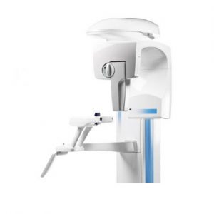
Easy upgrade from 2D to 3D
Planmeca ProMax® – future proof and a great investment
Planmeca ProMax® 2D is designed with upgradeability in mind. The unit’s modular structure allows easy conversion to different imaging modalities, while the software-driven SCARA is extremely flexible, allowing you to benefit from new imaging projections.
Whether you’re upgrading your 2D unit to 3D, or adding a cephalometric arm, Planmeca has the right solution for you. Individual options can be installed before delivery or added later, making Planmeca ProMax the most versatile all-in-one X-ray unit available.
Specifications
Imaging programs
| Standard: Basic panoramic programs | Standard panoramic Lateral TMJ (closed & open) PA TMJ (closed & open) PA sinus | |
| Standard | Child (Paediatric) mode for each standard and optional program to reduce the dose | |
| Optional | Horizontal and vertical segmenting for panoramic program | |
| Optional | Bitewing | |
Technical data
| Generator | Constant potential, resonance mode high frequency 80–150 kHz | |||||
| X-ray tube | D-054SB-P | |||||
| Focal spot size | 0.5 x 0.5 mm (IEC 336) | |||||
| Total filtration | min. 2.5 mm Al equivalent | |||||
| Anode voltage | 50–84 kV | |||||
| Anode current | 0.5–16 mA DC | |||||
| Exposure time | Pan | 2.7–16 s | ||||
| Scanning ceph | 6.4–9.9 s | |||||
| Planmeca ProCeph | 0.1–0.8 s | |||||
| Tomo | 3 s / frame | |||||
| SID | Pan | 500 mm (19 in.) | ||||
| Ceph | 163–170 cm (64–67 in.) | |||||
| Magnification | Pan | constant 1.2 | ||||
| Ceph | 1.08–1.13 | |||||
| CCD pixel size | 48 µm | |||||
| Image pixel size | 48/96/144 µm selectable | |||||
| CCD active surface | Pan | 6 x 147 mm | ||||
| Ceph | 6 x 295 mm | |||||
| Resolution (digital) | Pan | max. 9 lp/mm | ||||
| Ceph | max. 5.7 lp/mm | |||||
| Image field (digital) | Pan | 14 x 30 cm (5.5 x 12 in.) | ||||
| Ceph | 24/27 x 18/30 cm (9/10.6 x 7/11.8 in.) | |||||
| File size, uncompressed (digital) | Pan | 4–33 MB | ||||
| Ceph | 7–16 MB | |||||
| Line voltage | 100–240 V, 50 or 60 Hz | |||||
| Regulation | Automatic, ±10 % | |||||
| Line current | 8–16 A | |||||
| Colour | White (RAL 9016) | |||||
Physical space requirements
| Planmeca ProMax 2D | Planmeca ProMax 2D with cephalostat | ||||
| Width | 96 cm (38 in.) | 194 cm (76 in.) | |||
| Depth | 125 cm (49 in.) | 125 cm (49 in.) | |||
| Height* | 153–243 cm (60–96 in.) | 153–243 cm (60–96 in.) | |||
| Weight | 113 kg (lbs 248) | 128 kg (lbs 282) | |||
Minimum operational space requirements
| Planmeca ProMax 2D | Planmeca ProMax 2D with cephalostat | ||||
| Width | 150 cm (59 in.) | 215 cm (85 in.) | |||
| Depth | 163 cm (64 in.) | 163 cm (64 in.) | |||
| Height* | 243 cm (96 in.) | 243 cm (96 in.) | |||
*The maximum height of the unit can be adjusted for offices with limited ceiling space.
Dimensions